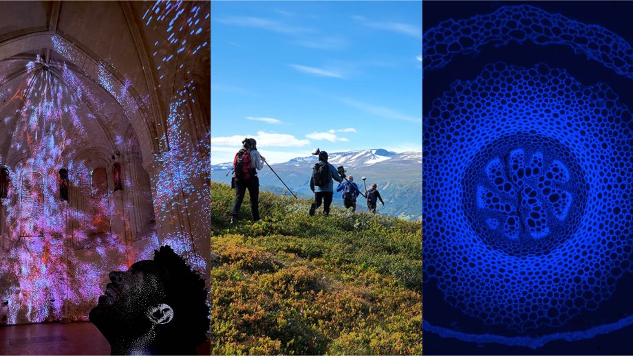
As researchers gain new ways of understanding the brain they hope to improve access to non-invasive ways of diagnosing and finding effective treatments for central nervous system disorders like schizophrenia, autism, and fetal alcohol spectrum.
To this end, postdoctoral scholar Dr. Sheryl Herrera (UWinnipeg/Cubresa) and Fulbright Scholar Maxina Sheft (Georgia Tech) have been working with University of Winnipeg physicist Dr. Melanie Martin on a study into how to identify anatomical changes in the brain.
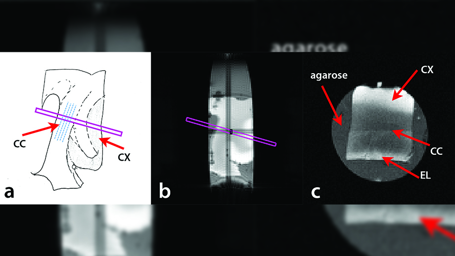
Human corpus callosum sample. (a) Drawing of the image slice taken for the experiment. The slice taken (long, thin box) was selected to be approximately perpendicular to the assumed direction of the axons (dashed lines) within the corpus callosum. (b) MR image of the slice taken from scanner to contain the corpus callosum (CC), ependymal layer (EL), and cortex (CX) regions. (c) MR image of the cross section taken from the MR slice from (b).
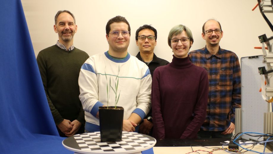
Herrera and Sheft are co-first authors on a recently published paper examining the first measurement of axon diameters in the human brain that measure only 2 one thousandths of a millimetre (2 μm). UWinnipeg alumni Dr. Morgan Mercredi (BSc, Honours, 2013), Dr. Richard Buist (University of Manitoba), Dr. Kanta Matsuda (Rutgers University), and Dr. Melanie Martin are also authors on this study.
The team’s findings are significant because previous axon measurements have typically been over 5 μm. By applying oscillating gradient spin echo (OGSE) sequences, the researchers were able to measure axons in the 1–2 μm range using a non-invasive imaging method. This capacity opens doors to long term brain studies that will increase understanding of how information is conducted.
Martin says that some of the most important axons in our brains are the smallest ones. These microscopic nerve fibres are several times thinner than human hair, yet they are vital to our ability to transmit information throughout our nervous system.
In a healthy brain, information is automatically processed and transmitted from the brain to different parts of the body. But in central nervous system disorders like schizophrenia there is evidence of abnormal function integration of brain processes. The more we can understand how this happens, the better chance we have of accurately diagnosing and finding treatments for these disorders.
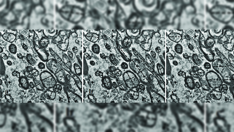
Measurements of axon diameters taken on Electron Microscope images. The dashed lines represent the lines drawn to collect the axon diameter measurements. (UWinnipeg)
Sheft was excited for the opportunity to use of a mathematical model to analyze the images and perform statistical analysis on its results to collect more accurate data.
“Improvements to our imaging methods can lead to better diagnostic methods at earlier stages, which is crucial for certain neurological disorders,” said Sheft. “The more accurate the images we can collect of the human brain, the more information we have to apply to diagnosing and treating various neurological disorders.”
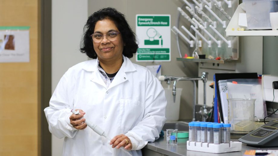
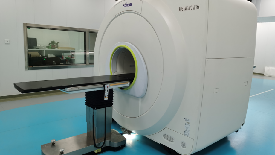
Both Sheft and Herrera were drawn to work in Martin’s lab because of the innovative work being done using MRI to diagnose central nervous system disorders. Sheft spent summer 2021 conducting research thanks to support from the Mitacs Globalink program. Herrera recently completed a Mitacs Elevate postdoctoral fellowship, which she says gave her unique access to MRI and PET technology to develop image acquisition and analysis techniques.
“I am applying my knowledge of the physics of MRI to a multidisciplinary approach to study the brain and its anatomical changes during central nervous system disorders like schizophrenia,” said Herrera. “I would love for this research to be utilized as a means for non-invasive diagnosis and treatment.”
This research was made possible thanks to a NSERC Discovery Grant, as well as Mitacs Elevate and Mitacs Globalink funding.